
Fig. 1.
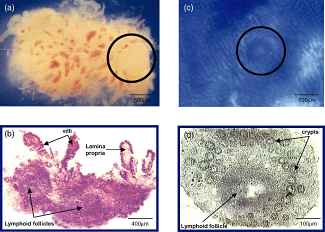
(a) Micrograph of an ileum biopsy (×20) containing a lymphoid follicle (circle); (b) histological confirmation (×25, haematoxylin and eosin stain). (c) Microscopic view of a colon sample after methylene blue staining (×20), the circle indicates a mucosal lymphoid follicle; (d) histological confirmation (×100).