
Fig. 2.
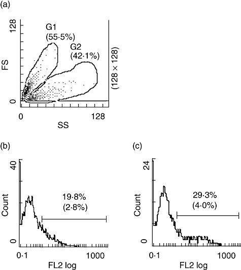
Complement activation on apoptotic Jurkat cells analysed by flow cytometry. Apoptotic cells were incubated with serum or with medium alone and analysed by flow cytometry. (a) Forward-scatter (FS) and side-scatter (SS) of apoptotic cells. Cells were gated into two regions, G1 containing early apoptotic cells and G2 containing late apoptotic cells. Results with apoptotic cells incubated with 10% normal human serum are shown. (b) Deposition of C3 in region G1 and (c) deposition of C3 in region G2 using antibodies against C3d. Percentages of positive cells are shown with percentages from control experiments within parenthesis. Histograms from one experiment typical of at least five are shown.