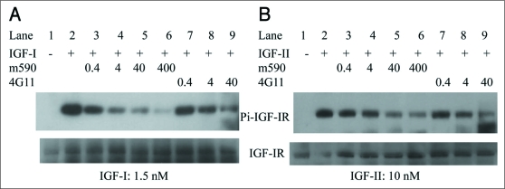
Figure 5.
Inhibition of IGF-I and IGF-II induced phosphorylation of IGF-IR by anti-IGF-IR human-mouse chimeric antibody IgG1 m590 and parental murine antibody IgG2b 4G11 in MCF-7 cells. MCF-7 cells starved in serum-free medium were pre-incubated with indicated concentrations (in nM) of IgG1 m590 (Lanes 3 to 6) and IgG2b 4G11 (Lanes 7 to 9) for 30 min followed by addition of 1.5 nM IGF-I (left) or 10 nM IGF-II (right) for 20 min. Total IGF-IR was immunoprecipitated with rabbit anti-IGF-IR beta subunit pAb. Phosphorylated IGF-IR was detected with mAb 4G10 specific to phosphotyrosine (top gels). The blots were re-probed with rabbit anti-IGF-IR beta subunit pAb (bottom gels) to show the total IGF-IR protein among the samples. Cells treated with IGF-I or IGF-II alone (Lane 2 in both panels) were included as positive control.

