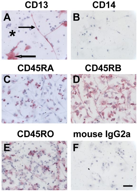
Figure 2. Expression of CD13, CD14, and CD45 isoforms on fibrocytes.
PBMC were cultured in SFM at 5×10⁵ cells/ml in 8-well glass slides for 7 days. Cells were then air-dried, fixed, and stained with antibodies. A) CD13, B) CD14, C) CD45RA, D) CD45RB, E) CD45RO, F) Irrelevant mouse IgG2a control. Cells were then counterstained with hematoxylin to identify nuclei. Positive staining was identified by red staining, with nuclei counterstained blue. Solid arrow points to a fibrocyte, open arrow points to a macrophage, and asterisk indicates a cluster of lymphocytes. Photomicrographs are representative results from at least four different donors. Bar is 50 µm.

