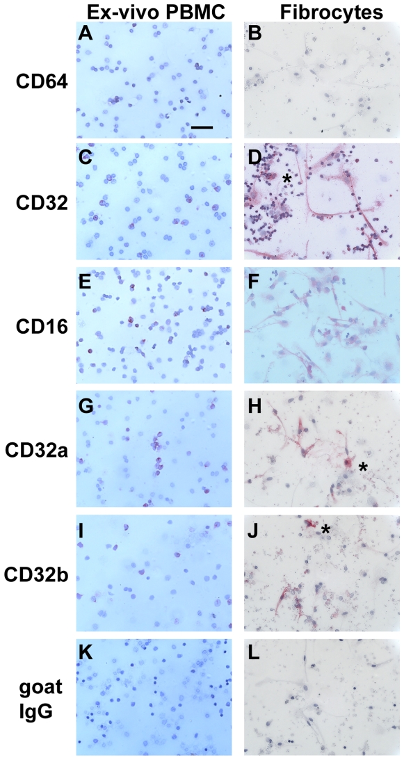
Figure 9. Comparison of Fc receptors on PBMC ex-vivo and fibrocytes.
PBMC were cultured in SFM at 5×10⁵ cells/ml in 8-well glass slides for 1 hour (ex vivo) or 7 days. Cells were then air-dried, fixed, and stained with antibodies against A and B) CD64, C and D) CD32, E and F) CD16, G and H) CD32a, I and J) CD32b, or K and L) goat IgG control antibodies. Cells were then counterstained with hematoxylin to identify nuclei. Asterisks are to the right of macrophages. Bar is 50 µm.

