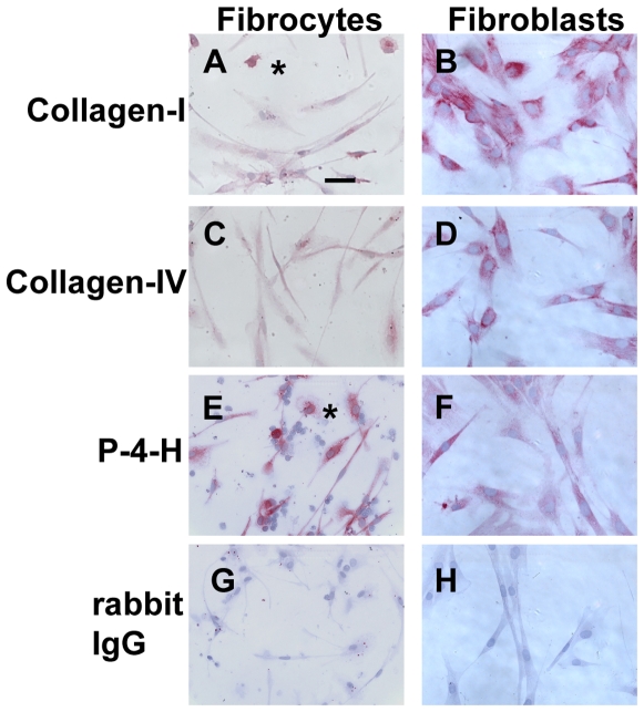
Figure 10. Comparison of collagen expression on fibrocytes and fibroblasts.
PBMC were cultured as described in Figure 1. Normal human dermal fibroblasts were cultured for 2 days in 8-well glass slides. Cells were then air-dried, fixed, and stained with antibodies. A and B) collagen-I, C and D) collagen IV, E and F) proyly-4-hydroxylase, G and H) rabbit IgG control antibody. Cells were then counterstained with hematoxylin to identify nuclei. Asterisks are to the right of macrophages. Bar is 50 µm.

