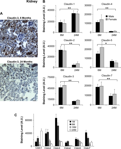
Figure 2.
Immunohistochemistry analysis of selected claudins in aging mouse kidney. (A) Representative staining for claudin-3 from kidney sections of 6- and 24-month-old male mice. Claudin-3 shows a significant decrease in older mice. (B) Effects of aging on the expression of the indicated claudins. Results shown were quantitated using the MCID image analysis software and represent the average of nine sections (three kidney punches from three mice) (*p < .05, **p < .001; brackets show the significance between age groups (both male and female) and the absence of a bracket indicates significance for one gender within the age group). The staining data are shown as arbitrary units (A.U.). Results are shown for 6- and 24-month-old mice. Black bars represent males and gray bars females. Claudin-2, -3, -4, -5, and -7 were significantly decreased in both male and female mice. Claudin-1 was increased in both males and females. (C) The effect of age and gender on claudin expression in mouse liver. The expression of the indicated claudins was examined in the liver of 1-month-old (1M), 6-month-old (6M), 16-month-old (16M), and 24-month-old (24M) mice. The same trends were observed when these additional time points were studied.

