Figure 1.

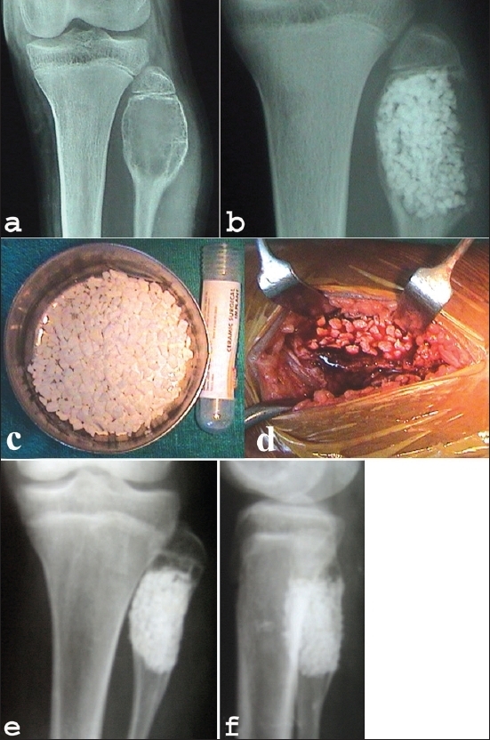
(a) Preoperative x-ray of knee AP view (a) shows a cavitary lesion (aneurysmal bone cyst) of upper end of fibula in 14 year old girl. Immediate postoperative x-ray (b) shows the cavity filled with HA granules. (c) and granule filled cavity (d) Peroperative clinical photograph of same patient showing HA granules (d). (e) Anteroposterior and lateral x-rays (f) of same patient at 2 yrs follow-up shows remodelling.
