
Figure 1A.
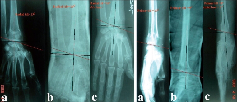
Pre-reduction AP x-ray (a) left wrist in dorsiflexion group shows fracture left radius with radial tilt of 15 degree. The radial tilt was corrected to 16 degree in postoperative xray (b) 6 months followup the xray (c) shows radial tilt of 16 degree. Overall no loss of radial tilt at 6 months followup. Fig. 1B - Pre-reduction lateral x-ray (a) of the same patient in dorsiflexion group shows palmar tilt of 10 degree which was corrected to 9 degree in postoperative xray (b) 6 months followup xray (c) shows palmar tilt of 8 degree. Overall 1 degree lost at 6 months followup