Figure 2.

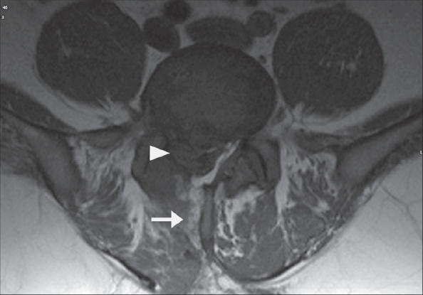
Axial MR image of a patient in the revision category - MRI showing track of previous procedure (arrow) and sequestered fragment in canal (arrow head)

Axial MR image of a patient in the revision category - MRI showing track of previous procedure (arrow) and sequestered fragment in canal (arrow head)