Figure 3.
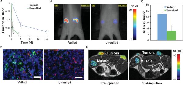
Effects of removable polymer coatings on the blood-clearance and tumor accumulation of nanoparticles. A) Nanoparticles bearing MMP cleavable polymer coatings (veiled) have greater blood clearance times compared with particles that have had the coating removed by MMPs (unveiled). Error bars indicate standard deviation of three animals. B) Fluorescence molecular tomography (FMT) of regions of interest (ROIs) selected around bilateral flank tumors from two representative animals shows intravenous injections of veiled nanoparticles yield greater accumulation in tumors after 48 h as compared to unveiled controls. C) Analysis of nanoparticle accumulation in the tumor at 48 h by FMT demonstrates superior accumulation of veiled particles as compared to unveiled controls. Error bars represent standard deviation of three animals. D) Representative histological sections confirm the increased accumulation of veiled nanoparticles versus unveiled controls after 48 h; nanoparticles (green), blood vessels (red), nuclei (blue). Scale bar is 250 μm. E) T2 map of tumor and muscle ROIs after intravenous injection show enhanced contrast from veiled nanoparticles in the tumor versus normal tissue (muscle) at 24 h post-injection.

