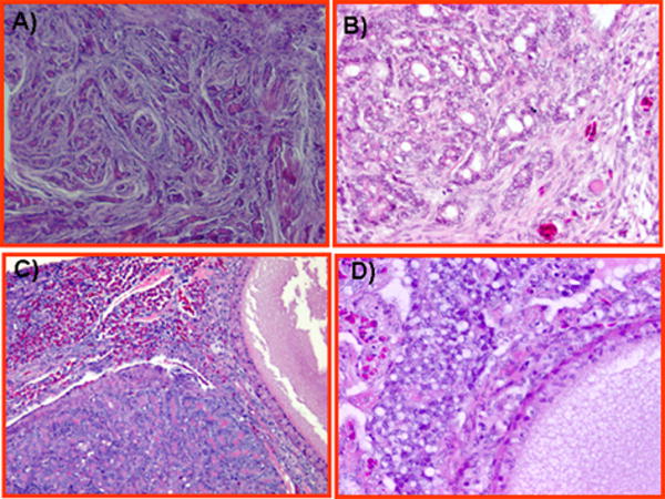
FIGURE 5.
Histological types of poorly differentiated ovarian epithelial carcinoma in hens. A, Poorly differentiated ovarian serous carcinoma showing solid areas composed of slitlike sheets containing cells with high-grade nuclear atypia. A few tiny glands are also seen without any papillae. B, Poorly differentiated ovarian endometrioid carcinomas showing complex glandular and microglandular patterns. Nuclear polymorphism, mitotic activities, and necrosis are marked. C, Poorly differentiated ovarian mucinous carcinomas showing confluent microglandular architecture in a cribriform pattern with a high grade of nuclear atypia and no intervening stroma. Moderate to strong eosinophilic reactions are also seen in the stroma. D, Poorly differentiated ovarian clear cell carcinoma showing vacuolated cells containing high-grade nuclear atypia that invade the stroma and theca layer of stromal follicles. Deposition of eosinophilic hyalinized matrix in the stroma and necrotic bodies are also seen. Hematoxylin and eosin, original magnification ×40.

