
FIGURE 7.
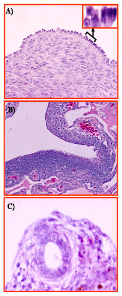
Figure 7. Putative precursor lesions of ovarian carcinoma in hens. A, Section of hen ovary with non-tumor abnormalities showing transformation of the ovarian surface epithelial layer from single columnar epithelial cells to a rounded phenotype with mitotic nuclei consistent with a malignant potential. Inset showing magnified (120×) view of transformed and normal surface epithelial cells. B, Section of hen ovary with non-tumor abnormalities. Marked epithelial dysplasia is seen in the surface layer with stromal invaginations. C, Section of hen ovary with non-tumor abnormalities showing inclusion cysts in the cortex beneath the ovarian surface epithelium containing epithelial cells of rounded phenotype. H &E, 40×.