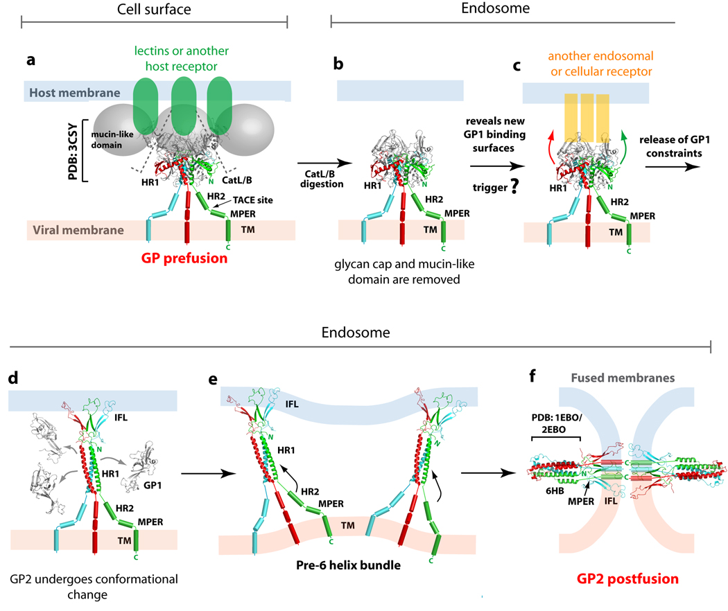
Figure 2. Ebolavirus GP-mediated entry.
Ebolavirus is thought to enter cells through receptor-mediated endocytosis (a) Initially, the metastable, prefusion EBOV GP may bind low affinity lectins or another unidentified receptor at the cell surface for viral attachment. (b) Subsequently, Ebolavirus is internalized and trafficked to the endosome, where host cathepsins cleave GP to remove the glycan cap and mucin-like domain. (c) The newly exposed surface may allow either tighter binding to the host surface receptor or binding to a second cellular factor or receptor in the endosome that could then trigger conformational changes in the GP2 fusion subunit. (d) Structural rearrangements in GP2 allow HR1 to form a single 44-residue helix and position the internal fusion loop for insertion into the host endosomal membrane. Upon insertion in the host membrane, the internal fusion loop adopts a 310 helix. (e) Based on studies in the influenza virus, more than one trimer of GP2 may be required during the membrane fusion process. (f) The formation of the low energy 6-helix bundle (6HB) requires HR2 and MPER to swing from the viral membrane towards the host membrane and pack against the trimeric bundle of HR1. These rearrangements juxtapose the EBOV GP’s internal fusion loop and transmembrane domain, thus facilitating the fusion of the host and viral membranes. This figure is adapted from [22].

