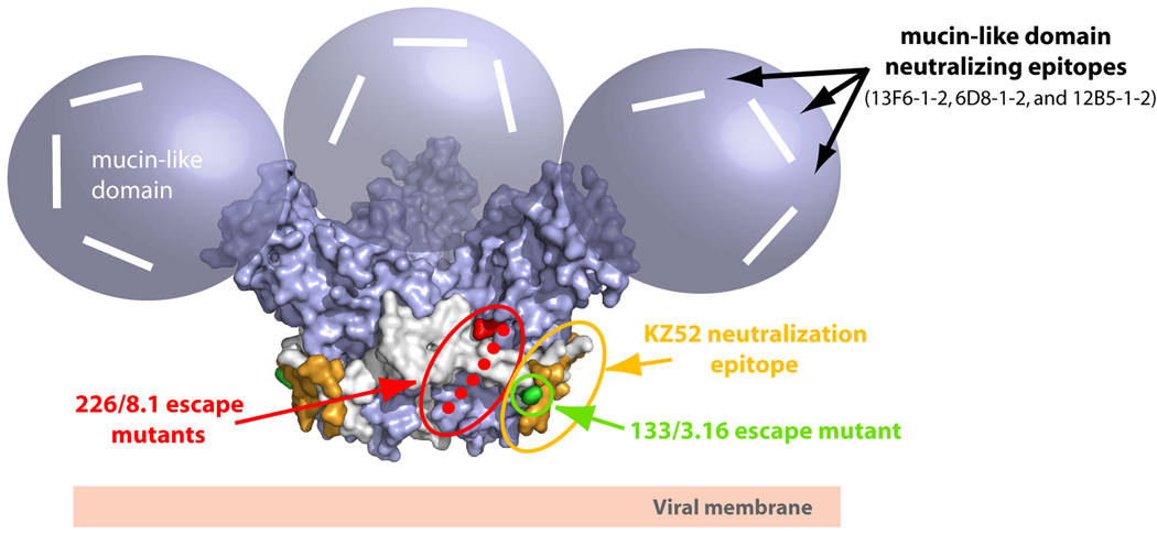
Figure 3. Locations of neutralizing epitopes on EBOV GP.
The locations of the Zaire ebolavirus neutralizing antibodies are mapped onto the molecular surface of the prefusion EBOV GP structure. In general, there are at least three regions on EBOV GP which have elicited neutralizing or protective epitopes. The KZ52 neutralizing epitope, which likely overlaps with mAb 133/3.16 (green), is located at a non-neutralizing site at the base of the EBOV GP chalice (colored in orange). This epitope is primarily composed of GP2 residues 505–514 and 549–556. A second neutralizing epitope termed 226/8.1 (colored red) is centered in the vicinity of the cathepsin cleavage site around residues 134, 194 and 195. The loop between residues 189–213 is disordered in the crystal structure and is shown as green dots. The mucin-like domain is the site of at least three linear neutralizing epitopes (modeled as white lines). These three linear neutralizing epitopes (residues 401–417, 389–405 and 477–493) map to unstructured and non-glycosylated regions on the mucin-like domain.

