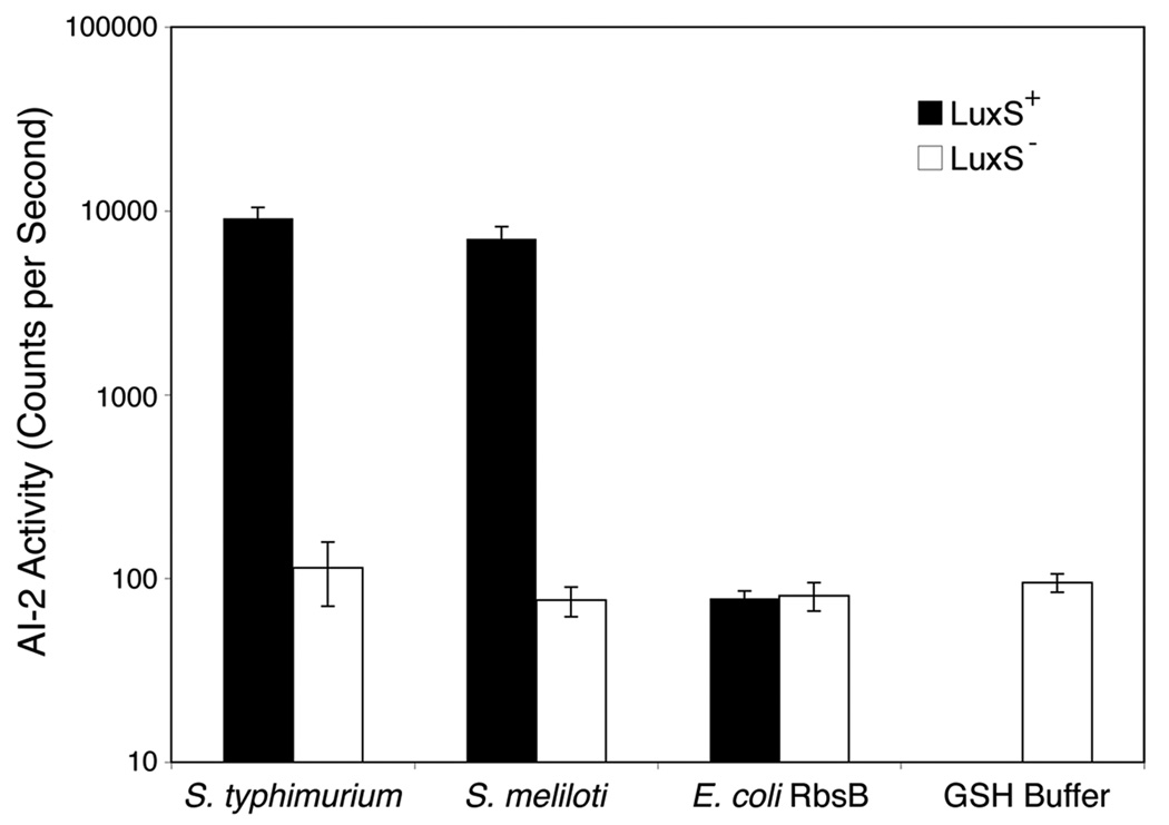
Fig. 2. Binding of AI-2 to potential receptor proteins.
Light produced by V. harveyi strain MM32 (LuxN−, LuxS−) was assayed following the addition of ligand released from purified protein expressed in either LuxS+ (black bars) or LuxS− (white bars) E. coli (strains BL21 and FED101, respectively). The E. coli ribose binding protein RbsB and protein-free GSH-buffer were included as negative controls. AI-2 activity is reported as c.p.s. of MM32 bioluminescence. Error bars represent the standard deviations for three independent cultures.

