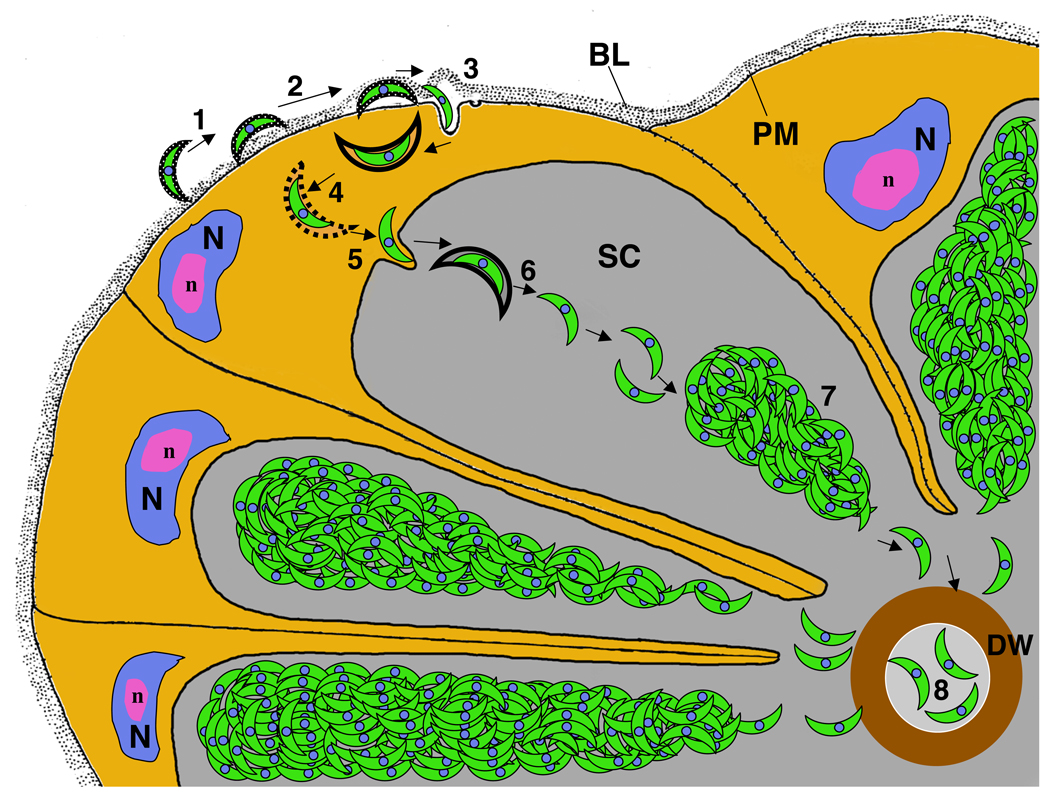
Figure 2. Progression of sporozoite invasion of the salivary gland.
1) The sporozoite attaches to the basal lamina; 2) Sporozoite passage to the space between the basal lamina and the basal epithelial cell plasma membrane, a process associated with the loss of the sporozoite’s thick coat; 3) Penetration of the basal plasma membrane; the sporozoite resides within a vesicle; 4) Release of the sporozoite from the surrounding membrane by an unknown mechanism; 5) Invasion of the apical membrane and entry into the secretory cavity; 6) Sporozoites are released from the surrounding membrane by an unknown mechanism; 7) Sporozoites assemble into bundles within the secretory cavity; and 8) A small number of sporozoites enter the secretory duct by an unknown mechanism. BL: Basal Lamina; DW: Duct wall; N: nucleus; n: nucleolus; PM: Plasma membrane; SC: Secretory cavity. Modified from reference [•5].

