
Figure 1.
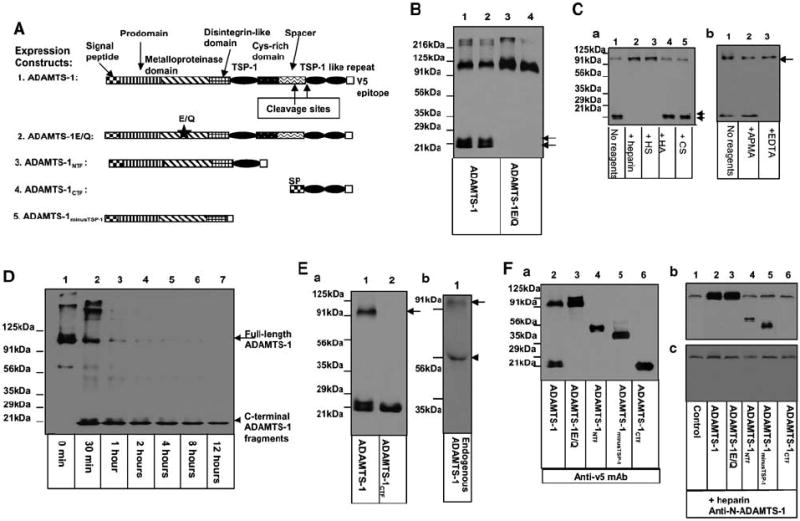
ADAMTS-1 undergoes auto-proteolytic cleavage, and the self-cleavage of ADAMTS-1 is regulated. (A) Diagram of the expression constructs used in the experiments. (B) The proteolytic cleavage of ADAMTS-1 requires its own metalloproteinase activity. The cell culture supernatants derived from Cos-7 cells transfected with the COOH-terminal v5-epitope-tagged ADAMTS-1 (lanes 1 and 2) or ADAMTS-1E/Q (lanes 3 and 4) were analysed by Western blotting with anti-v5 antibody. Arrows indicate the COOH-terminal cleavage fragments of ADAMTS-1. (C) The cleavage of ADAMTS-1 is inhibited by heparin and HS. In (Ca), TA3ADAMTS-1 cells were cultured in the absence (lane 1) or presence of 100 μg/ml of heparin (lane 2), HS (lane 3), hyaluronan (lane 4), or CS (lane 5) for 48 h. The COOH-terminal cleavage fragments are indicated by arrows. In (Cb), TA3ADAMTS-1 cells were cultured in the absence (lane 1) or presence of 200 μm APMA (lane 2) or 500 μm EDTA (lane 3) for 48 h. Cell supernatants were analysed by Western blotting with anti-v5 antibody. (D) Auto-proteolytic cleavage capacity of ADAMTS-1 was assessed by incubating 100 ng of purified ADAMTS-1 in Tris buffer at 37°C for 0 min (lane 1), 30 min (lane 2), or 1 (lane 3), 2 (lane 4), 4 (lane 5), 8 (lane 6), or 12 (lane 7) h. Reaction products were assessed by Western blotting with anti-v5 antibody. (Ea) Western blotting with anti-v5 mAb shows that v5-tagged ADAMTS-1CTF (lane 2) displays a molecular size similar to that of the smaller COOH-terminal cleavage fragment of ADAMTS-1 (lane 1). (Eb) Western blotting of supernatants derived from TA3wt cells using a polyclonal rabbit antibody against an NH2-terminal peptide of mouse ADAMTS-1 (anti-N-ADAMTS-1). (F) Supernatants were derived from TA3wtb (lane 1), TA3ADAMTS-1(lane 2), TA3ADAMTS-1E/Q (lane 3), TA3ADAMTS-1NTF (lane 4), TA3ADAMTS-1minusTSP-1 (lane 5), or TA3ADAMTS-1CTF (lane 6) cells that were cultured in the absence (Fa) or presence (Fb and c) of 100 μg/ml of heparin. Supernatants were analysed directly by Western blotting with anti-v5 antibody (Fa) to assess the expression levels of the v5-tagged exogenous ADAMTS-1 variants or with anti-N-ADAMTS-1 polyclonal antibody (Fb) to detect both endogenous and exogenous ADAMTS-1 and its NH2-terminal variants. One set of supernatants were passed through anti-v5 antibody affinity columns to absorb the v5-tagged exogenous ADAMTS-1 variants. Equal amounts of the flow-through proteins were analysed by Western blotting with anti-N-ADAMTS-1 antibody to detect endogenous ADAMTS-1 (Fc).