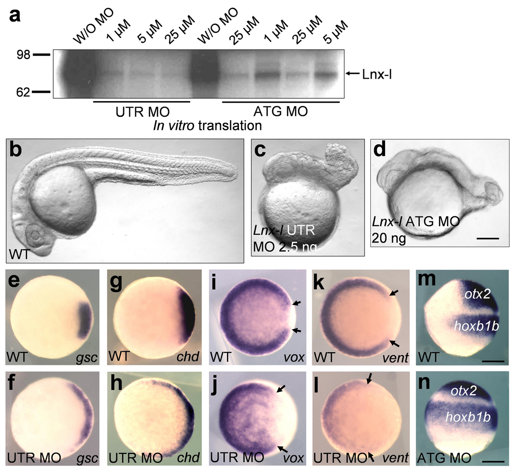
Figure 1. Depletion of Lnx-l causes dorsalization.

a, Both MOs inhibit in vitro translation of lnx-l mRNA; lnx-l UTR MO is more efficient than ATG MO. b–d, lnx-l UTR MO (2.5 ng) and ATG MO (20 ng) both caused similar embryonic defects displaying degenerated tail and trunk; 26 hpf embryos, lateral views. e–l, lnx-l morphants (UTR MO, 2.5 ng) show ventro-laterally expanded expression of gsc (f) and chd (h), and reduced expression of vox (j) and vent (l), with arrows indicating enlarged dorsal gap in the vox and vent domains; germ ring stage, animal pole views. m and n, lnx-l ATG MO (20 ng) injected embryos showed extended expression of otx2 and hoxb1b; 80% epiboly stage, lateral view, dorsal is to the right. Scale bar, 200 µm.
