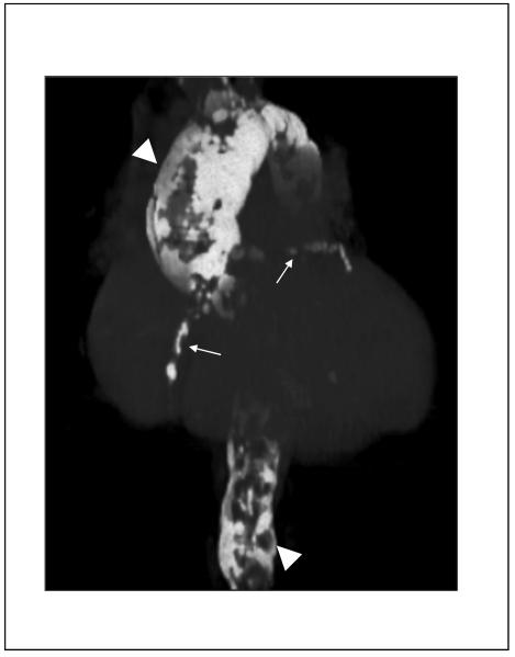
Figure 5.
Computed tomogram of severe aortic and coronary calcification in a patient with a remote history of aortitis (“porcelain aorta”). Shown are volume-rendered CT images demonstrating severe calcification of the thoracic and abdominal aorta (arrowheads), as well as the coronary arteries (arrows). There is diffuse aneurysmal dilatation of the ascending thoracic aorta. (Image courtesy Dr. Paul Schoenhagen, Departments of Radiology and Cardiovascular Medicine, Cleveland Clinic Foundation).

