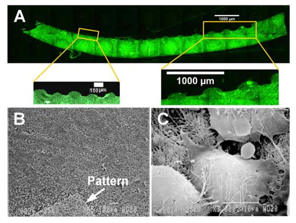
Figure 3.

(A) Fluorescent image of the cross-section of a patterned CG membrane perpendicular to the channel direction, showing the morphological characteristics of the patterned features. The insets show the wide and narrow width channels at higher magnification. (B) SEM image of bovine aortic endothelial cells (BAEC's) seeded onto a sample patterned CG flow channel network (the pattern is marked by an arrow) (C) SEM image of an individual cell with extension of processes and establishment of cell-cell junctions
