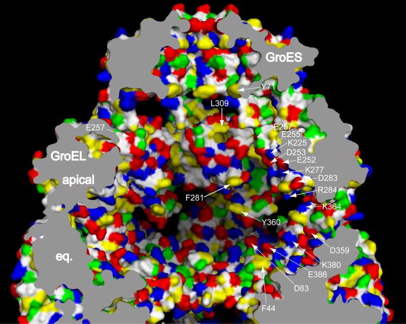
Fig. 2. Cis GroEL/GroES folding chamber.
The domed chamber formed by GroES binding to a nucleotide-bound GroEL ring is shown in a cutaway view of a space-filling model, with the wall character revealed by coloring of amino acid side chains: red, acidic; blue, basic; yellow, hydrophobic; green, polar; white, main chain. Parts of three subunits are visible. A number of residues are identified by the arrowing. Note that there is considerable electrostatic character to the wall (see text). eq., equatorial domain. Figure adapted from ref.22.

