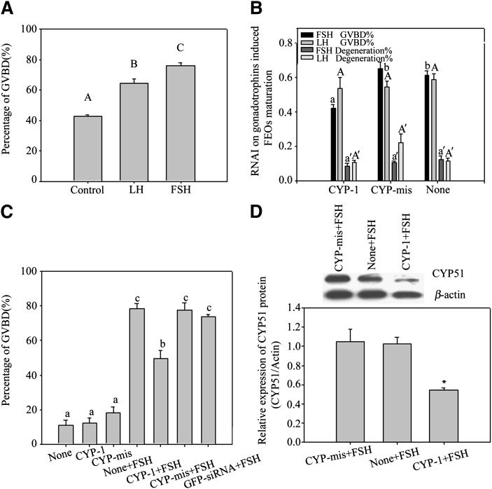
Fig. 4.
A, B: FEOs in vitro maturation after gonadotrophin induction with or without RNAi treatment. In A, both FSH (more than 70%) and LH (more than 60%) remarkably stimulated FEO maturation in 12 h (P < 0.05), but the LH effect was weaker than FSH. Bars with different capital letters show significant differences (P < 0.05). In B, during the 12 h culture period, siRNAs moderately decreased the GVBD rate induced by FSH (43% in CYP-1 vs. 66% in control, P < 0.05), in which about 23% of FEO meiotic maturation was inhibited from maturation. Instead, LH induced FEO maturation was unaffected (P > 0.05) (58% in CYP-1 vs. 63% in control, P > 0.05). The rate of oocyte degeneration was also presented in B and oocyte degeneration was about 20% in 12 h culture. Different letters on the same colored bars show significant difference (P < 0.05), i.e., letter a only compare with b, a’ only compare with b’, A only compare with B, A’ only compare with B’. C, D: The effects of RNAi on FSH-induced CEO in vitro maturation. In C, the influence of RNAi on CEO maturation. Twelve h after siRNAs transfection, CEOs were cultured in media with or without 10 U/l FSH and further cultured for 12 h. The oocyte maturation results showed that CYP-1 significantly prevented FSH stimulated CEOs meiotic resumption, as the GVBD ratio data proved (P < 0.05). Different lowercase letters on the bars show significant differences, n = 3 (P < 0.05). In D, Western blot of FSH-induced CYP51 expression in CEOs after RNAi. Twelve h after transfection, CEOs were transferred into media containing 10 U/l FSH and further cultured for 8 h. The results show observably decreased CYP51 protein levels in CEOs. This figure represents one of the similar results in three independent experiments. The groups with an asterisk (*) show significant difference compared with None + FSH group, n = 3 (P < 0.05).

