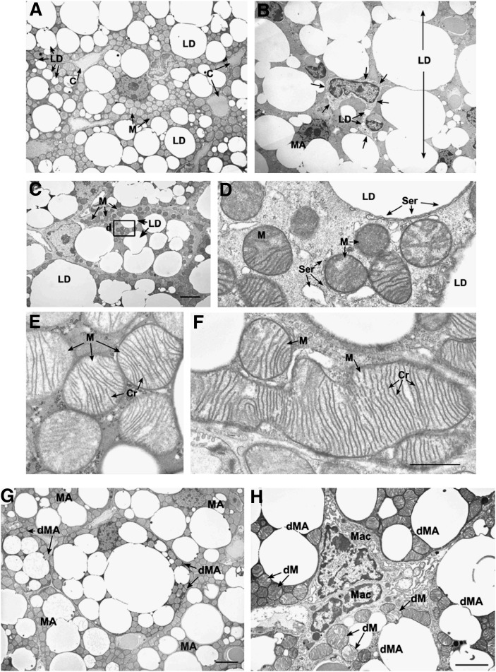
Fig. 6.
Influence of Pctp expression on the ultrastructure of brown fat. The top series of electron micrographs show that the absence of Pctp expression leads to the presence of immature adipocytes and mitochondrial changes in brown fat. Characteristics of brown fat from wild-type (A, E) or Pctp−/− mice (B, C, D, F). A: Mature brown adipocytes in wild-type mice. LD, lipid droplet; C, blood capillary; M, mitochondria. B: Developing protoadipocyte at the same magnification as in A. LD, lipid droplet (two in protoadipocyte and one large fused large droplet); MA, mature adipocytes. Arrows mark the boundary of the protoadipocyte. C: Developing preadipocyte at the same magnification as in A. LD, lipid droplet; M, mitochondria. Bar in C applies to A–C and represents 4 µm. D: High magnification of the area in the box in C. Note that smooth endoplasmic reticulum (Ser) is found along the edge of the lipid droplet (LD), surrounding mitochondria (M), and in the cell cytoplasm. E: Mitochondria from control adipocytes (in A). Note that mitochondria (M) have a pale matrix and numerous cristae (Cr) that are well organized. F: Mitochondria from Pctp−/− mouse that is the same magnification as in E. Note the large size of mitochondria (M) and the large number of parallel cristae (Cr). Bar in F applies to D–F and represents 1 µm. The bottom pair of electron micrographs demonstrates that degenerating adipocytes are present in brown fat from Pctp−/− mice. G: Degenerating mature adipocytes (dMA) were found among mature adipocytes (MA) in the brown fat from Pctp−/− mice. The cytoplasm of degenerating brown adipocytes is electron dense in comparison to mature adipocytes. Bar represents 4 µm. H: The intercellular space between degenerating mature adipocytes (dMA) in brown fat from Pctp−/− mice is expanded, and macrophages (Mac) can be found in close association with the degenerating cells. In addition to electron-dense cytoplasm in degenerating mature adipocytes, mitochondria have an abnormal structure with disrupted cristae, pale matrix, and defects in shape (dM). Bar represents 6 µm.

