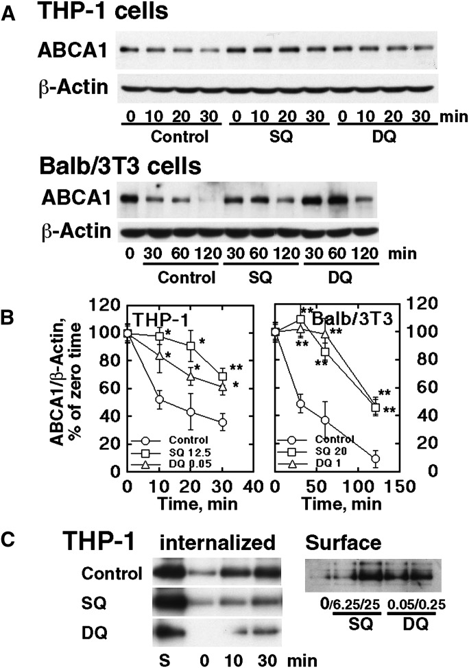
Fig. 2.
Stabilization of ABCA1 by SQ and DQ. A: Retarded degradation of ABCA1 in the presence of cycloheximide in THP-1 macrophages and Balb/3T3 cells, by SQ (25 nM for THP-1 and 20 nM for Balb/3T3) and DQ (0.05 nM and 1.0 nM). B: Graphic expression of the results typically represented by Fig. 2A after standardization for β-actin. Error bars indicate SE for three measurements. Significant difference from control at each time point is indicated as * P < 0.05 and ** P < 0.01. C: Internalization of ABCA1. Left panel: THP-1 cells were preincubated with SQ (25 nM) and DQ (0.25 nM) for 16 h to equilibrate the cells with the compounds. The surface ABCA1 was then labeled by biotinylatin and the cells were incubated for time indicated. The surface biotinylation was cleaved and the remaining biotinylated ABCA1 was analyzed as the protein internalized. Right panel: Cell surface ABCA1 was analyzed by surface biotinylation after incubation with SQ and DQ (as indicated in nM) for 1 h.

