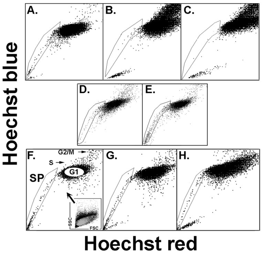
FIGURE 1.
Presence of SP cells in cultured conjunctival epithelium as a function of time in culture and medium. Rabbit cells after 16 (A), 40 (B, D), and 64 (C) hours of culture in SHEM or 40 hours in base medium (E). Human cells after 16 (F), 40 (G), and 64 (H) hours in SHEM. (F) Includes a description of the light-scatter gate used to exclude nonepithelial cells from the SP and nSP cohorts examined in the microarray studies. The Hoechst plot SP, G1, S, and G2/M are indicated.

