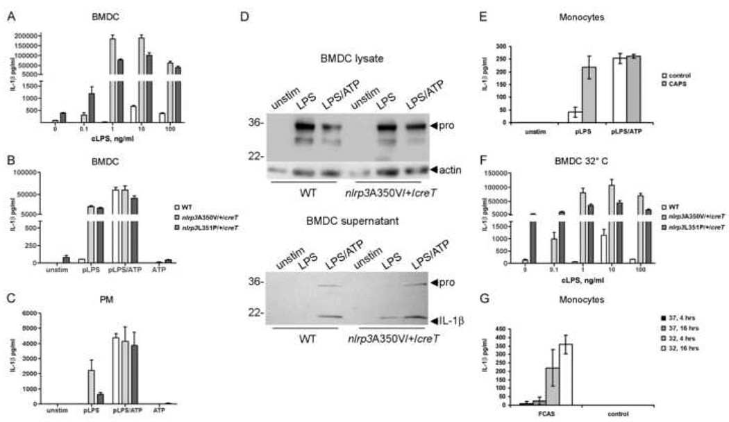
Figure 3. Myeloid cells from Nlrp3A350V/+/CreT and Nlrp3L351p/+/CreT mice and CAPS patients react similarly to innate stimuli.
Tamoxifen-treated Nlrp3A350V/+/CreT, Nlrp3L351P/+/CreT, and littermate WT BMDC were incubated with varying amounts of crude LPS (A) or pure LPS (100 ng/ml) with and without ATP (5mM) (B). (C) Tamoxifen-treated peritoneal macrophages (PM) were incubated with pure LPS with and without ATP. IL-1β in the supernatants was measured by ELISA. (A–C) n=2–3 mice with 3 wells each / genotype. (D) Western blotting for IL-1β or actin loading control, Nlrp3A350V/+/CreT BMDC lysates (top), and supernatants (bottom). (E) In vitro stimulation of monocytes from 3 CAPS patients and 3 normal human controls with pure LPS with and without ATP. (F) In vitro cold stimulation of WT, Nlrp3A350V/+/CreT, and Nlrp3L351P/+/CreT BMDC, n=2–3. Cells were incubated at 32°C overnight and supernatants were analyzed by ELISA for IL-1β. (G) In vitro cold stimulation of monocytes from 5 FCAS patients and 2 normal human controls, 4 hrs and overnight. Also shown, incubation at 37°C.

