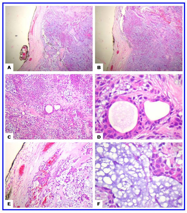
Figure 2.
The histological features of the juvenile pleomorphic adenoma of the cheek. (a-b) Scanning magnification views of pleomorphic adenoma showing that the tumor is well circumscribed. The mesenchymal and epithelial elements are evident at these magnifications. This encapsulated tumor has a protuberance pushing through the capsule (upper left side). The tumor has abundant chondroid areas. (c-d) These views show the essential cytoarchitectural features sufficient for diagnosis of pleomoprhic adenoma. The tubular strucutes are well delineated from the stroma. They are lined by inner layer of ductal epithelial cells and other layer of myoepithelial cells. The latter merge imperceptivity into the surrounding mesenchymal elements. The tubules have eosinophilic colloid like materials (secretions) in their lumens. The myoepithelial cells appear as cuboidal, spindle, plasmacytoid and non-descript epithelioid cells. They surround the tubules in a thin layer and in focally thick mantles. (e-f) These views show the stroma (one of the defining component of the pleomorphic adenoma). It is composed of hyaline, homogenous, fibrillary (e) and chondromyoxoid (f) materials interspersed among the epithelial and myoepithelial cells.

