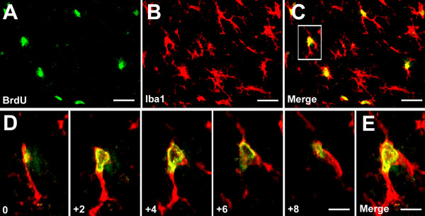
Figure 4.
Confocal microscopy images show BrdU expression in spinal microglia two days after SNI. (A-C) Colocalization of BrdU and Iba1 in the medial superficial spinal cord. Scale bars, 50 μm. (D) Stack of confocal images (2 μm apart) from a double-labeled cell enlarged from a square in C. (E) Merge of all images in D. Scale bars, 20 μm.

