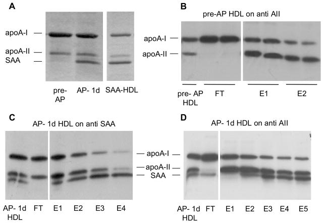
Figure 3.
Apolipoprotein characterization of pre-AP HDL, AP- 1d HDL and SAA-HDL. (A) SDS gel of HDL (5 μg total protein). (B) 125I-pre-AP HDL (5 μg) was passed through an anti-apoA-II immunoaffinity column and fractions were electrophoresed and autoradiographed as outlined in the methods (ARG). (C) 125I-AP HDL (10 μg) was passed through an anti-SAA immunoaffinity column with subsequent electrophoresis and ARG as described in (B). (D) 125I-AP HDL (10 μg) was passed through an anti-apoA-II immunoaffinity column with subsequent electrophoresis and ARG as described in (B). Note: the gels in B–D were loaded on the basis of 2000 cpm per lane and since the majority of counts were present in E1 and E2, E3–E5 quantitatively represent a smaller percentage of total protein mass.

