
Figure 4.
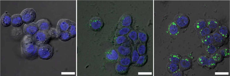
Overlayed DIC and confocal fluorescence images of the DRAQ5 channel (blue, nuclear stain) and the BDC-NH-BODIPY channel (green) of HT-29 cells incubated with no particles (a), 0.19 mg/mL of 1b@silica particles (equivalent to 17 μM BODIPY) (b), and 0.38 mg/mL of 1b@silica particles (equivalent to 34 μM BODIPY) (c). The bars represent 25 μm.