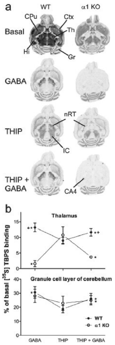
Fig. 3.

Modulation of [35S]TBPS binding by GABA and THIP in α1 KO mice. a, Representative images of [35S]TBPS binding and its modulation by 10 mM GABA, 1 mM THIP or both on wild-type and α1 KO mouse horizontal brain sections. CA4, CA4 area of the hippocampus; Ctx, cortex; CPu, caudate-putamen; Gr, granule cell layer of the cerebellum; Hi, hippocampus; nRT, reticular nucleus of the thalamus; Th, thalamus; IC, inferior colliculus. All the binding was abolished in the presence of 100 μM picrotoxinin as in Figs 1 and 2 (not shown). The representative images were scanned and processed using identical scaling for brightness and contrast, but the basal binding image was taken from films exposed only for 2 days rather than 21 days. b, Quantitative proportions of the [35S]TBPS binding in the presence of 10 mM GABA, 1 mM THIP or both in the thalamus and granule cell layer of cerebellum. Data are means ± SEM (n = 5 for α1 KO and n = 6 for wild-type mice) and expressed as percent of the corresponding basal [35S]TBPS binding value. *p < 0.05 for the significance of the difference between GABA and THIP or between THIP and THIP + GABA (paired t-test). + p < 0.05 for the significance of the difference from the corresponding wild-type value (Bonferroni t-test).
