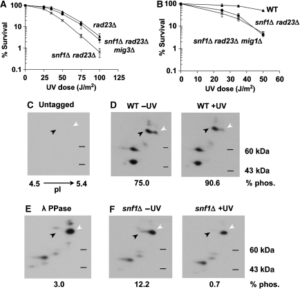
Figure 5.
Mig3 is a downstream target of Snf1. (A and B) Effect of mig3Δ and mig1Δ on the UV sensitivity of snf1Δ rad23Δ cells. UV survival curves were obtained as in Figure 3A. (C–F) Two-dimensional gel electrophoresis and western blotting of whole cell extracts from untagged (C) or Mig3-myc containing (D-F) cells. Samples were taken from WT (D) or snf1Δ cells (F) before and 5 min after UV irradiation, as indicated. Myc-tagged Mig3 migrates with an apparent molecular weight of 70 kDa on a standard one-dimensional denaturing protein gel. Two major species of Mig3-myc consistent with this molecular weight were detectable, and are indicated by the black (phosphorylated) and white (unphosphorylated) arrows. Loss of phosphorylation is evident in lambda phosphatase-treated WT sample (E).

