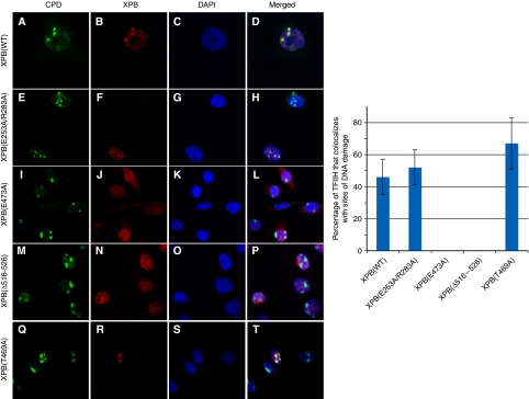
Figure 5.
Recruitment of TFIIH to local sites of DNA damage. (Left panel) CHO-27-1 cells were stably transfected with pEGFP plasmids expressing various forms of GFP-tagged XPB proteins. These cells were irradiated with UV light (100 J/m2) through the 3-μm pore filter and fixed 30 min later. Immunofluorescent labelling was performed using either a mouse monoclonal anti-CPD (panels A, E, I, M, Q) or a rabbit polyclonal anti-GFP (panels B, F, J, N, R). Nuclei were counterstained with DAPI (panels C, G, K, O, S), and slides were merged (panels D, H, L, P, T). (Right panel) Quantitative analysis of the recruitment of TFIIH to sites of DNA damage in transfected cells. Values represent averages±s.d. (n=100 sites of DNA damage) from three independent experiments.

