Figure 1.
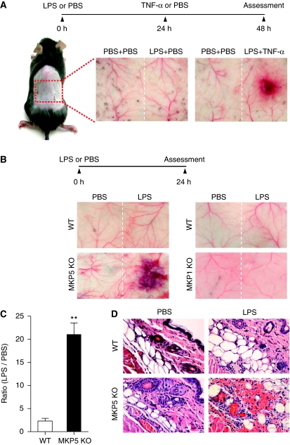
LPS-induced microvascular injury in Mkp5+/+ and Mkp5−/− mice. (A) Experimental scheme and results of a classical local Shwartzman reaction (LSR) induced by consecutive injections of LPS and then TNF-α. Dorsal skin of WT mice were first injected s.c. with LPS (80 μg, right side of each panel) or PBS control (left side). After 24 h, TNF-α (0.2 μg) or PBS in same volume was injected s.c. into the same site that received LPS. The mice were killed 24 h after the second injection, and the skin tissues were examined macroscopically. Representative sample images from one of the five experiments are shown. (B) Experimental scheme and results of a modified (one-injection) LSR showing macroscopic appearance of dorsal skin in Mkp5+/+ (WT) and Mkp5−/− (KO) mice (panels on the left), compared with that of the Mkp1+/+ (WT) and Mkp1−/− (KO) mice (panels on the right). The WT and KO littermates in each group received an injection of either LPS (80 μg; right to the dotted line) or PBS (left to the dotted line). A total of 11 WT and 15 Mkp5−/− mice, and 8 WT and 8 Mkp1−/− mice were examined 24 h after the LPS injection. Representative images are shown. (C) The degree of haemorrhage in the Mkp5+/+ and Mkp5−/− group above was estimated based on densitometry analysis of skin samples receiving either LPS or PBS injection. **P<0.01. (D) Grouped images showing representative photomicrographs of H&E-stained skin sections from WT (upper panels) and Mkp5−/− (lower panels) mice that were treated with LPS or PBS as marked. Erythrocyte extravasation, thrombus formation and neutrophil accumulation are evident in the sample from LPS-treated Mkp5−/− mice 24 h after LPS injection.

