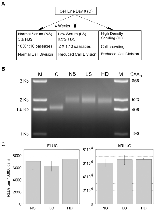
Figure 4. GAA·TTC expansion is independent of cell division rates.
(A) To analyze the influence of cell division on GAA·TTC expansion, a parental cell line was cultured under various conditions affecting cell-division rates: Normal Serum (NS, 5% FBS growth media) in which cells were passaged 10 times over the 4 week time period; Low Serum (LS, 0.5% FBS growth media) in which cells were passaged only twice over the 4 week time period due to reduced cell division; High Density Seeding (HD) cells were plated and carried near confluent cell levels in normal growth media in order to reduce cell division due to crowding. (B) PCR analysis of a (GAA·TTC)352 insert from a cell line cultured for 4 weeks in various conditions affecting cell-division rates. C is the Day 0 control. PCR amplification adds 438 bp to the GAA·TTC insert. M: 1 Kb plus size standard. A representative gel from an n = 3 is shown. (C) Analysis of the effects of the different growth conditions on transcription levels through the integrated reporter constructs. Analysis of the FLUC and hRLUC reporter expression levels is shown. Expression is represented as relative light units (RLUs) per 40,000 cells. Error bars represent the standard error of the mean (SEM) from an n = 3.

