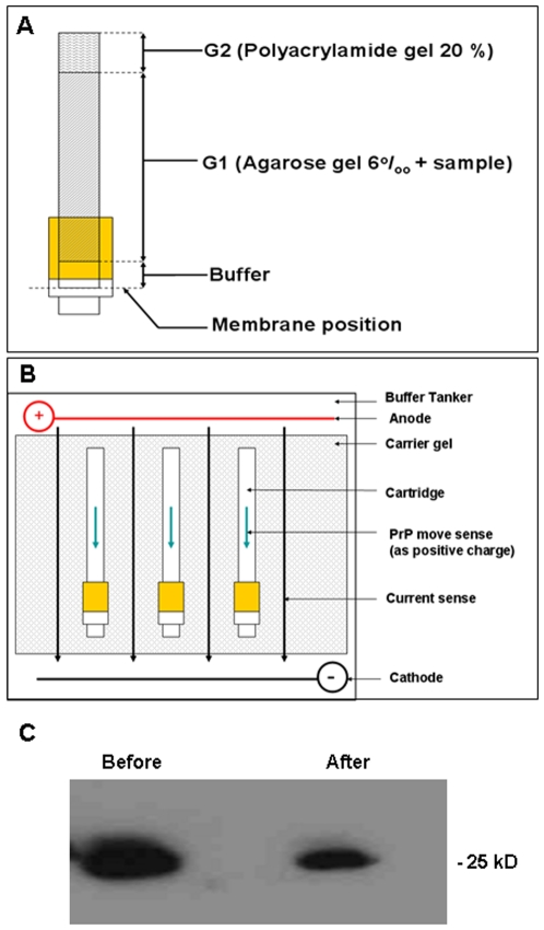
Figure 3. Electrophoretic Desorption.
A Schematic diagram of the desorption device. The cartridge with the membrane at the base is loaded with the components as indicated. The agarose component is mixed with the sample and added over the buffer when the cartridge is in the upright position. Once the agarose has solidified the polyacrylamide plug is added. B The cartridge with the components added is laid flat in a standard DNA electrophoresis chamber. Buffer is then added to cover the cartridge and current applied. C Western blot of recombinant PrP detected with and antibody 8B4. The protein was the same molecular weight before and after absorption and desorption from the mte clay matrix and retained its N-terminus.

