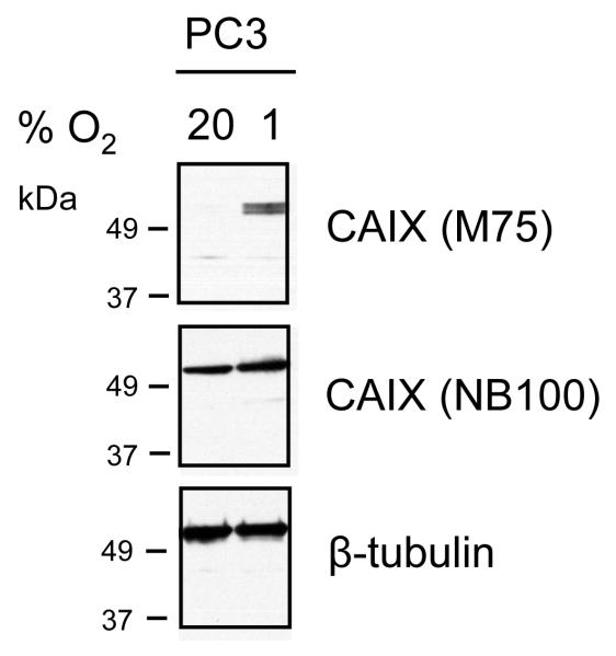
Figure 3. Expression of CAIX and tubulin in PC-3 prostate cancer cells.
PC-3 cells were cultured under normoxic or hypoxic conditions (1% O2 for 48 hours). Cell lysates (15ug) were separated on 10% SDS-PAGE gels and transferred to nitrocellulose membranes. Blots were probed with antibodies to tubulin (1:1000) and CAIX (NB100 and M75, both at 1:5000 dilution).

