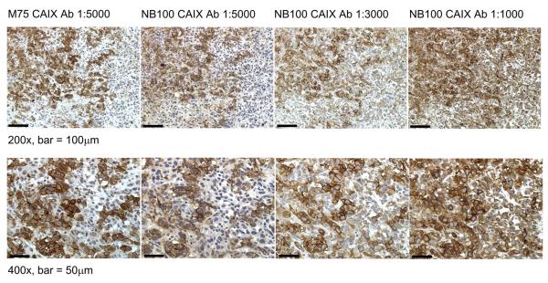
Figure 4. Detection of CAIX in xenograft tumors grown from PC-3 cells in mice.
Mice were injected subcutaneously in the flank with 3×10⁶ PC-3 prostate cancer cells and tumors were resected on Day 39. Immunohistochemical staining of Formalin-fixed, paraffin-embedded sections was performed using antibodies to CAIX (NB100 at 1:1000, 1:3000 and 1:5000 dilutions; M75 at 1:5000 dilution). Antigen retrieval was performed by incubating slides in 10 mM citric acid buffer pH 6.0, as described in Methods. Stained sections were photographed at using a Zeiss Axioplan 2 imaging microscope and Openlab 5.0.3 Beta Improvision® software.

