
Figure 8.
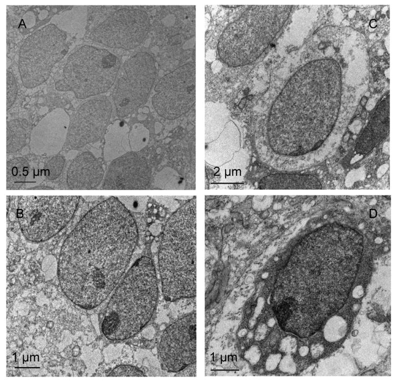
TEM pictures of human embryonic stem cells on flexible mat: After day 2: close cell-cell contacts throughout aggregates (A, B), After day 10: separation of cells by pericellular ECM components (C), assuming oval ‘chondron’-like structures, abundant fragmented collagen fibers (D)