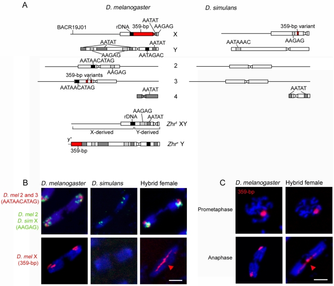
Figure 2. Separation failure of the D. melanogaster X chromatids during anaphase in hybrid female embryos.
(A) Schematic of satellites and other loci used as targets of FISH, and of Zhr mutant and duplicated chromosomes. Mapping of FISH probes is shown in Figure S2. Boxes and lines represent heterochromatin and euchromatin, respectively. Constrictions in boxes represent the centromeres. Chromosomal regions are not drawn to scale. The Zhr 1 chromosome is thought to have resulted from a recombination between an X and a Y [25]. Our FISH analysis shows that this compound-XY chromosome contains the Y centromere and the proximal half of the Y long arm fused with the X euchromatin and distal pericentric heterochromatin (Figure S2); note that this structure differs from that inferred by Sawamura et al. [25]. In this chromosome, the entire 359-bp satellite block has been deleted. The Zhr + Y chromosome was made by a translocation onto the distal end of the Y long arm of half of the X-derived 359-bp satellite block and a small amount of distal euchromatin carrying the y + marker from an inverted X chromosome (In(1)sc 8) [36]. (B) Top row, D. melanogaster 2 and 3 chromatids and the D. simulans X chromatids segregate normally in hybrid female embryos. Bottom row, the D. melanogaster X chromatids, marked by a probe for the 359-bp satellite (red arrowhead), fail to segregate and comprise the lagging chromatin. (C) The 359-bp satellite block is normally condensed during prometaphase but abnormally stretched (red arrowhead) during anaphase in hybrid female embryos. DNA is blue in (B) and (C). Scale bar is 5 µm in (B) and (C).

