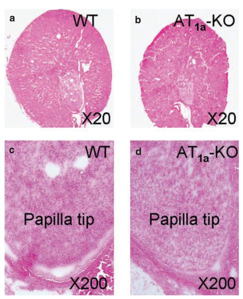
Figure 1. Anatomical and low-power microscopic visualization of cortical and inner medullary structures of WT and AT1a-knockout (KO) mice.
(a) A representative midline cross section of a WT kidney. (b) A representative midline cross section of an AT1a-KO kidney. (c) A representative low-power micrograph of a WT kidney showing the papilla tip of the inner medulla. (d) A representative low-power micrograph of an AT1a-KO kidney showing the papilla tip of the inner medulla. Kidney sections were stained with hematoxylin and eosin. WT, wild type.

