
Figure 1.
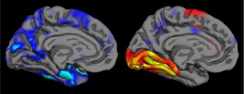
The localization, magnitude, and extent of abnormalities observed in fMRI studies of patients with neurologic diseases depend on both localization and severity of pathology and on functional networks engaged by the particular fMRI task, as well as participant performance on the task. In this illustration, regions of cortical thinning in Alzheimer’s disease from structural MRI (left, (Dickerson et al., 2007b)) are compared with cortical areas activated, as measured with fMRI, in normals during an event-related study of successful learning of new information that was able to later be freely recalled (right, (Dickerson et al., 2005a)). Analytic tools are emerging that enable the direct investigation of relationships between functional and structural abnormalities in MCI/AD and other disorders. Figure used with permission. (WE NEED TO REQUEST PERMISSION FROM NeuroRx, Elsevier—Dickerson BC, July 2007)