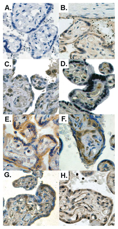
Figure 4. Examples of immunohistochemistry staining.

The expression and tissue distribution of the different hypoxia markers were assessed on fixed placental tissue sections. A. The specificity of the staining was checked by omitting the primary antibody. B. PlGF syncytial and endothelial staining of a malaria-infected placenta. C. Flt-1 positive cells are found in the syncytium, villus vessel walls and Hofbauer cells. D. KDR staining is mainly found in the villus vessels endothelium. In control placentas (E.), VEGF mainly stained the syncytium but malaria-infected placentas with intervillositis, (F.), showed significantly more frequent and intense staining of stromal cells. However, no difference was found for HIF-1α staining when uninfected placentas (G.) were compared with malaria-infected ones (H.), both showing syncytial and stromal staining (possibly Hofbauer cells). Original magnification: 200x. Colour balance, luminosity and contrast have been optimised.
