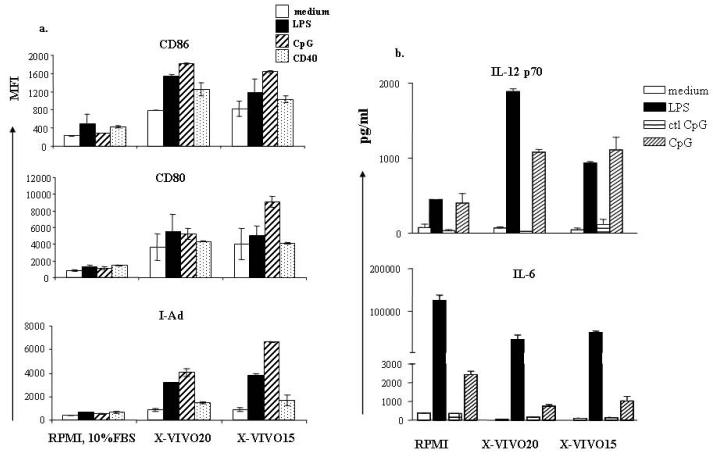
Figure 2. Expression of costimulatory molecules and pro-inflammatory cytokine of serum-free cultured DCs.
DCs were grown in different culture media as described. DCs were further stimulated as indicated on day 6 for 48 hours (a) or 16-18 hours (b). (a) DCs were exposed to inflammatory stimuli, washed with PBS, and stained with fluorescence conjugated antibodies to CD11c, CD80, CD86, and I-Ad. MFI of CD80, CD86, and I-Ad was calculated from CD11c-positive cells exclusively. (b) DCs were plated with inflammatory stimuli indicated in 24 well plates at 106/ml for 16-18 hours. Supernatants were harvested and cytokines were measured by Sandwich ELISA. Results represent mean concentration ± SD of 5 individual experiments.

