
Figure 1.
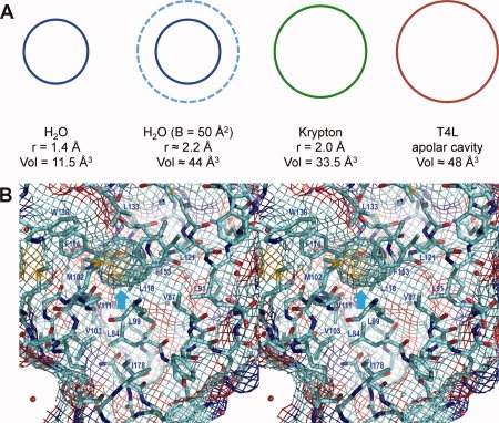
A: Sketch comparing, from left to right, the volume occupied by a water molecule at rest, a water molecule with a thermal factor of B = 50 Å2, a krypton atom at rest and an apolar cavity present in wildtype T4 lysozyme. At a pressure of 8bar, krypton is seen to bind within the lysozyme cavity (occupancy ∼0.4; Quillin et al., 2000), even though there are no direction-specific interactions to help localize the krypton atom. Crystallographic analysis suggests that the occupancy of water in this cavity is very low. B: Stereo pair showing the apolar cavity present in pseudo-wildtype lysozyme (WT*; C54T/C97A; PDB code 1L63). The “chicken wire” grid shows not only the internal cavity (cyan arrow) but also the external (solvent-exposed) surface of the protein. If, for example, there is an oxygen atom (red) close to the cavity, the surface grid at that location is also shaded red. This figure as well as Figures 2(B) and 3 were prepared with PyMOL (DeLano, 2008).