
Figure 3.
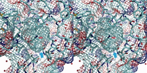
Stereo pair showing the cavity in the mutant T4 lysozyme L99A/M102L (PDB code 3DKE). As in Figure 1(B), the “chicken wire” grid shows both the internal cavity (cyan arrow) and the external solvent-exposed surface of the protein. The site of the Leu99 to Ala substitutions is shown in red. Most of the cavity wall is colored turquoise, indicating its nonpolar nature. Close to the carbonyl group of Ala99, however, the red color indicates some polar character. There is also some dark blue and red in the vicinity of Val87.