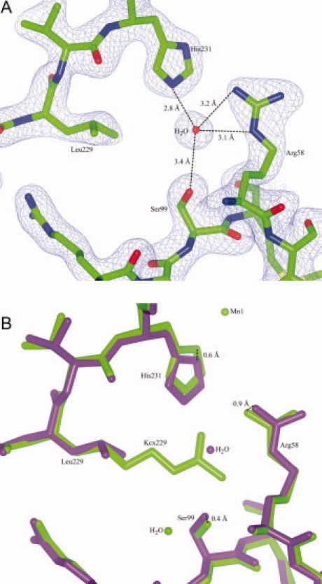
Figure 5.

Structure of UVDE K229L. Panel A: Detail of the model and the map of UVDE K229L showing the environment of Leu229. Distances of the water molecule to neighboring residues are indicated. Panel B: Detail of a superposition of UVDE K229L (in dark purple) with the original structure of UVDE (in light green). The slight shifts of residues Arg58, Ser99, and His231 are indicated. [Color figure can be viewed in the online issue, which is available at www.interscience.wiley.com.]
