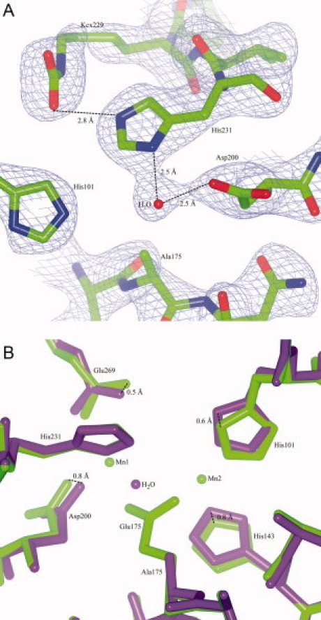
Figure 6.

Structure of UVDE E175A. Panel A: Detail of the map (contoured at 1.25 σ) and the model of UVDE E175A showing the environment of Ala175 and the presence of a carboxylated lysine. Distance between an oxygen atom of Kcx229 and His231 is indicated as well as the distances to the water taking the place of Mn1 and Mn2 to neighboring residues. Panel B: Detail of the superposition of the original structure of UVDE (in light green) and UVDE E175A (in dark purple) showing the surroundings of residue Glu175/Ala175. [Color figure can be viewed in the online issue, which is available at www.interscience.wiley.com.]
