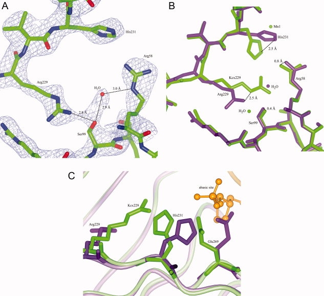
Figure 7.

Structure of UVDE K229R and model with DNA. Panel A: Model and map (contoured at 1.5 σ) of UVDE K229R. Distances between a water molecule and Arg58 and Ser99 are indicated. Panel B: Detail of the superposition of the original structure of UVDE (in light green) and UVDE K229R (in dark purple) showing the position of Arg229 compared with the carboxylated lysine and the large shift in the position of His231. Also the shifts in the position of Arg58 and Ser99 are indicated. Panel C: Detail of superposition of the original structure of UVDE (in light green) and the structure of UVDE K229R (in dark purple) in which DNA with an abasic site was modeled (orange) based on a comparison with the structure of endonuclease IV with damaged DNA. Arg229/Kcx229, His231, and Glu269 are depicted in cylinder representation, the abasic site of the DNA is depicted in ball-and-stick representation whereas the rest of the protein and the DNA is depicted in ribbon representation. [Color figure can be viewed in the online issue, which is available at www.interscience.wiley.com.]
